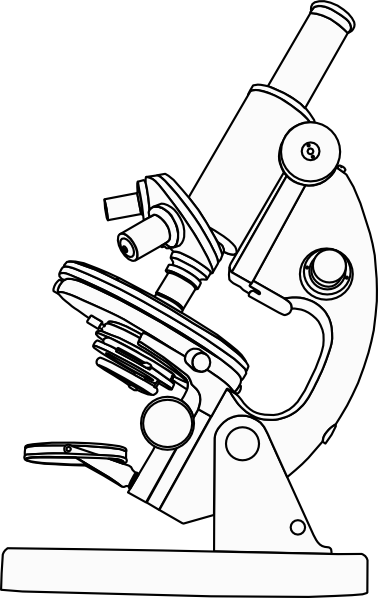
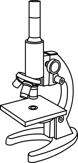
39 compound microscope diagram without labels
Fluorescence - Wikipedia The chemical compound responsible for this fluorescence is matlaline, which is the oxidation product of one of the flavonoids found in this wood. [1] In 1819, Edward D. Clarke [5] and in 1822 René Just Haüy [6] described fluorescence in fluorites , Sir David Brewster described the phenomenon for chlorophyll in 1833 [7] and Sir John Herschel ... Inorganic Chemistry 4th edition, Catherine Housecroft Enter the email address you signed up with and we'll email you a reset link.
Looking at the Structure of Cells in the Microscope ... A special sample holder is used to keep this hydrated specimen at -160°C in the vacuum of the microscope, where it can be viewed directly without fixation, staining, or drying. Unlike negative staining, in which what is seen is the envelope of stain exclusion around the particle, hydrated cryoelectron microscopy produces an image from the ...
Compound microscope diagram without labels
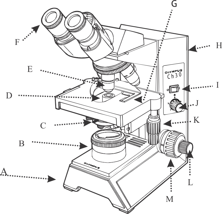
Microscope Labeling - The Biology Corner Students label the parts of the microscope in this photo of a basic laboratory light microscope. Can be used for practice or as a quiz. Microscope Labeling Game - PurposeGames.com About this Quiz. This is an online quiz called Microscope Labeling Game. There is a printable worksheet available for download here so you can take the quiz with pen and paper. This quiz has tags. Click on the tags below to find other quizzes on the same subject. Science. Diagram of a Compound Microscope - Biology Discussion A bright-field or compound microscope is primarily used to enlarge or magnify the image of the object that is being viewed, which can not otherwise be seen by the naked eye. Magnification may be defined as the degree of enlargement of the image of an object provided by the microscope.
Compound microscope diagram without labels. What is a Compound Microscope? - New York Microscope Company A compound microscope is an instrument that is used to view magnified images of small specimens on a glass slide. It can achieve higher levels of magnification than stereo or other low power microscopes and reduce chromatic aberration. It achieves this through the use of two or more lenses in the objective and the eyepiece. A Study of the Microscope and its Functions With a Labeled Diagram ... Compound Microscope Diagram The compound microscope uses light for illumination. Some compound microscopes make use of natural light, whereas others have an illuminator attached to the base. The specimen is placed on the stage and observed through different lenses of the microscope, which have varying magnification powers. Compound Microscope Parts, Functions, and Labeled ... Compound Microscope Definitions for Labels. Eyepiece (ocular lens) with or without Pointer: The part that is looked through at the top of the compound microscope. Eyepieces typically have a magnification between 5x & 30x. Monocular or Binocular Head: Structural support that holds & connects the eyepieces to the objective lenses. Solved Label the image of a compound light microscope using - Chegg Expert Answer. 100% (17 ratings) Transcribed image text: Label the image of a compound light microscope using the terms provided.
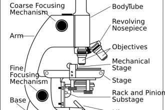
Compound Microscope: Parts of Compound Microscope - BYJUS (A) Mechanical Parts of a Compound Microscope 1. Foot or base It is a U-shaped structure and supports the entire weight of the compound microscope. 2. Pillar It is a vertical projection. This stands by resting on the base and supports the stage. 3. Arm The entire microscope is handled by a strong and curved structure known as the arm. 4. Stage Simple Microscope - Parts, Functions, Diagram and Labelling A compound microscope is also called a bright field microscope. It can provide magnification by up to 1,000 times. Stereo microscope/dissecting microscope - It can magnify objects by up to 300 times. It is used to visualize opaque objects that cannot be visualized using a compound microscope. The Compound Light Microscope - Miami University The microscope pictured above is referred to as a compound light microscope. The term light refers to the method by which light transmits the image to your eye. C ompound deals with the microscope having more than one lens. Microscope is the combination of two words; "micro" meaning small and "scope" meaning view. CODEX multiplexed tissue imaging with DNA-conjugated ... - Nature Jul 02, 2021 · A newly adapted version of CODEX uses an automated microfluidics system and conventional fluorescent microscope to iteratively hybridize, image and strip fluorescently labeled DNA probes that are ...
How to Use a Compound Microscope: 11 Steps (with Pictures) - wikiHow Focus the microscope. Looking through the eyepiece, arrange the illuminator and the diaphragm to reach the most comfortable level of light. Move the specimen slide so that the image is in the center of your view. [10] Arrange the illuminator until you've arrived at a comfortable level of light. Binocular Microscope Anatomy - Parts and Functions with a Labeled Diagram Now, I will discuss the details anatomy of the light compound microscope with the labeled diagram. Why it is called binocular: because it has two ocular lenses or an eyepiece on the head that attaches to the objective lens, this ocular lens magnifies the image produced by the objective lens. Binocular microscope parts and functions Compound Microscope - Types, Parts, Diagram, Functions and Uses A compound microscope captures an inverted image of the specimen because every time the light passes through the lens, the image's direction is flipped. The image always ends up inverted from the original. So, if you move the sample to the left, it moves in the right direction. Image 18: A comparison image between a simple and compound microscope. Compound Microscope: Basics, Functionality, and Uses The word compound means multiple, mix, or a combination of both. A compound microscope is a microscope which uses more than one lens. Devised with a system of combination of lenses, a compound microscope consists of two optical parts, namely the objective lens and the ocular lens. The objective lens uses short focal length to enlarge an image.
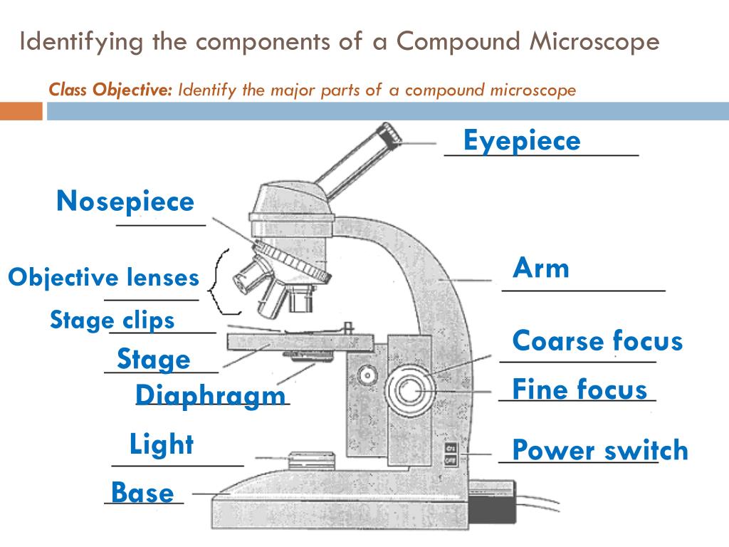
PPT - Identifying the components of a Compound Microscope PowerPoint Presentation - ID:2045335
Microscope Parts and Functions Microscope Parts and Functions With Labeled Diagram and Functions How does a Compound Microscope Work?. Before exploring microscope parts and functions, you should probably understand that the compound light microscope is more complicated than just a microscope with more than one lens.. First, the purpose of a microscope is to magnify a small object or to magnify the fine details of a larger ...

Microscope With Labels Clip Art at Clker.com - vector clip art online, royalty free & public domain
Label the microscope — Science Learning Hub In this interactive, you can label the different parts of a microscope. Use this with the Microscope parts activity to help students identify and label the main parts of a microscope and then describe their functions. Drag and drop the text labels onto the microscope diagram.

8 Best Images of Lens Diagram Worksheet - Microscope with Labeled Parts, Label Eye Parts ...
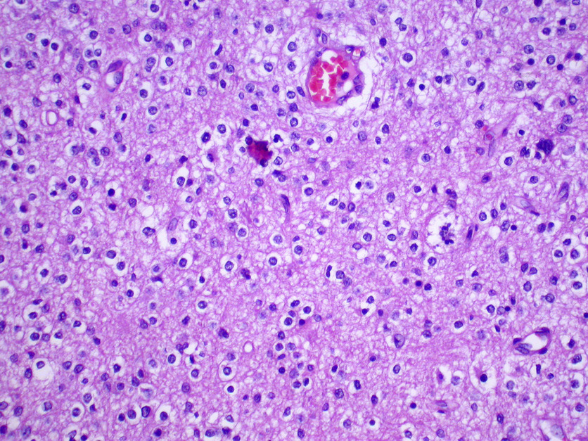
Lower Secondary Science LEARNER’S BOOK 8 - Issuu Feb 22, 2021 · The photograph in Figure 1.2.1 shows a tiny part of the lungs, seen through a powerful microscope. You can see the lungs are mostly holes. These holes are called air sacs.

(i) Draw a neat labelled diagram of a compound microscope explain briefly its working (ii) Why ...
Microscope Diagram Labeled, Unlabeled and Blank Print a microscope diagram, microscope worksheet, or practice microscope quiz in order to learn all the parts of a microscope.
Biology 521 Resources
16 Parts of a Compound Microscope: Diagrams and Video Once you have an understanding of the parts of the microscope it will be much easier to navigate around and begin observing your specimen, which is the fun part! The 16 core parts of a compound microscope are: Head (Body) Arm Base Eyepiece Eyepiece tube Objective lenses Revolving Nosepiece (Turret) Rack stop Coarse adjustment knobs
animal: Animal Cell Labeled Edgenuity
Electron microscope - Wikipedia An electron microscope is a microscope that uses a beam of accelerated electrons as a source of illumination. As the wavelength of an electron can be up to 100,000 times shorter than that of visible light photons , electron microscopes have a higher resolving power than light microscopes and can reveal the structure of smaller objects.

Eckhard Bick - PDF Free Download
Parts of a Compound Microscope and Their Functions - NotesHippo Compound microscope magnification is determined by multiplying the eyepiece and objective powers. When viewed through a 5X eyepiece with a 10X objective, an item is magnified 5 x 10=50 times. The magnification is 10 x 45 = 450 times when using a 10X eyepiece and a 45X objective. How to Use the Compound Microscope
Microscope Clip Art at Clker.com - vector clip art online, royalty free & public domain
Working Principle and Parts of a Compound Microscope (with Diagrams) It holds the stage, body tube, fine adjustment and coarse adjustment. 5. Body Tube: It is usually a vertical tube holding the eyepiece at the top and the revolving nosepiece with the objectives at the bottom. The length of the draw tube is called 'mechanical tube length' and is usually 140-180 mm (mostly 160 mm). 6.

Compound Microscope: Definition, Diagram, Parts, Uses, Working Principle
Compound Microscope- Definition, Labeled Diagram, Principle, Parts, Uses Alternatively, the magnification of the compound microscope is given by: m = D/ fo * L/fe where, D = Least distance of distinct vision (25 cm) L = Length of the microscope tube fo = Focal length of the objective lens fe = Focal length of the eye-piece lens Parts of a Compound Microscope Eyepiece And Body Tube.
Science : student_compund_microscope_outline_220 : Classroom Clipart
PDF Parts of a Microscope Printables - Homeschool Creations typical student microscope -other microscopes will vary) •Which part of the microscope rotates so another person can look through the eyepiece without needing to move the microscope ? the head •What is the magnification level on the eyepiece of a microscope?10x (see objective lens magnification to see how these work together)
Microscope Coloring
Labeling the Parts of the Microscope Labeling the Parts of the Microscope This activity has been designed for use in homes and schools. Each microscope layout (both blank and the version with answers) are available as PDF downloads. You can view a more in-depth review of each part of the microscope here. Download the Label the Parts of the Microscope PDF printable version here.

Diagram Of Liver Cell : Ib Biology Sl Eukaryotic Cells Flashcards Quizlet / As long as 25 ...
Compound Light Microscope: Everything You Need to Know A compound light microscope is a type of light microscope that uses a compound lens system, meaning, it operates through two sets of lenses to magnify the image of a specimen. It's an upright microscope that produces a two-dimensional image and has a higher magnification than a stereoscopic microscope. It also goes by a couple of other names ...
Get Practice Labeling Parts Of A Microscope Gif - DirectScot
Compound Microscope Parts Head/Body houses the optical parts in the upper part of the microscope. Base of the microscope supports the microscope and houses the illuminator. Arm connects to the base and supports the microscope head. It is also used to carry the microscope. When carrying a compound microscope always take care to lift it by both the arm and base ...

29 best Anatomy and Physiology images on Pinterest | Physiology, Anatomy and Anatomy reference
Microscope, Microscope Parts, Labeled Diagram, and Functions Revolving Nosepiece or Turret: Turret is the part of the microscope that holds two or multiple objective lenses and helps to rotate objective lenses and also helps to easily change power. Objective Lenses: Three are 3 or 4 objective lenses on a microscope. The objective lenses almost always consist of 4x, 10x, 40x and 100x powers. The most common eyepiece lens is 10x and when it coupled with ...

Q: Parts of a Compound Microscope
Compound Microscope Parts – Labeled - Rs' Science The term "compound" refers to the microscope having more than one lens. Basically, compound microscopes generate magnified images through an aligned pair of the objective lens and the ocular lens. In contrast, "simple microscopes" have only one convex lens and function more like glass magnifiers.
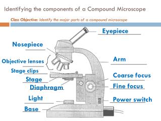
PPT - Identifying the components of a Compound Microscope PowerPoint Presentation - ID:2045335
Labelled Diagram of Compound Microscope The below mentioned article provides a labelled diagram of compound microscope. Part # 1. The Stand: The stand is made up of a heavy foot which carries a curved inclinable limb or arm bearing the body tube. The foot is generally horse shoe-shaped structure (Fig. 2) which rests on table top or any other surface on which the microscope in kept.

Compound Microscope Diagram - ClipArt Best
Parts of Stereo Microscope (Dissecting microscope) – labeled ... If you would like to learn optical components of a compound microscope, please visit Compound Microscope Parts – Labeled Diagram and their Functions, and this article. How to use a stereo (dissecting) microscope. Follow these steps to put your stereo microscopes in work: 1. Set your microscope on a tabletop or other flat sturdy surface where ...
Dissecting Microscope Labeled Diagram - Micropedia
Parts of a microscope with functions and labeled diagram - Microbe Notes Figure: Diagram of parts of a microscope There are three structural parts of the microscope i.e. head, base, and arm. Head - This is also known as the body. It carries the optical parts in the upper part of the microscope. Base - It acts as microscopes support. It also carries microscopic illuminators.
Post a Comment for "39 compound microscope diagram without labels"